Monday- Friday, Feb 15-19
While we had some previous lectures on iron gall ink in my Masters year, these past two weeks was spent on a more in-depth practice on the treatment of iron gall ink objects and on creating experiments involving iron gall inks. This workshop time spanned the two weeks between February 15th-February 26th.
In preparation of the workshop we prepared samples of different papers with different iron gall inks on them, which we then 'aged' in the oven (at 120°C for 10-20 minutes). The first week started with online lectures from Birgit Riessland (RCE) on Monday and with a workshop with Claire Phan-Tan-Luu (Practice-in-Conservation & Nationaal Archief) and Eliza Jacobi (Practice-in-Conservation & Rijksmuseum van Oudheden) on Tuesday.
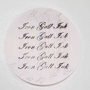
During the workshop we were able to see for ourselves some of the effects that some adhesives have on the distribution of Fe(II) ions in paper (mind you, on filter paper, so some of the worst case spreading). We made some pre-coated tissues for ourselves, to use on the samples we made over the weekend. We also tested different adhesives by using pre-coated tissues on another filter test paper.
We spent Wednesday and Thursday preparing some possible experiments to try out concerning iron gall ink and involving the aging chambers (further described at the bottom of the blog). These would be put into aging chambers the following week.
Friday morning was spent in a lecture with Han Neevel (RCE) on the science behind calcium phytate treatments while the afternoon was spent working more with Claire and Eliza.
Monday-Friday, Feb 22-26
This was the second week of the Iron Gall Ink workshop, spent applying the things we had learned the previous week and attempting a calcium pyhtate treatment. Monday we prepared the solutions for the phytate treatment and gathered our prepared iron gall ink samples that we had created and 'aged' the week before.
On Tuesday we attempted the treatment, running into some difficulties with the pH which we later concluded was the result of too much calcium present in the water we used from the very beginning of the preparation (leaving the solution to be slightly too alkaline to create a successful calcium phytate treatment). While we spent the rest of the week attempting the phytate solution and trying to find the reason for our pH issues, we also were attempting mends on the remaining 'aged' ink samples we had created. On Friday we collected all our findings at that point in order to present to the lecturers and supervisors.
A Small Experiment
In order to learn more about how humidity can affect the degradation of iron gall ink objects, we split up into groups and created samples to then be placed into aging chambers for two weeks. Cynthia and I decided to focus our samples on the amount of ink placed on a paper sample and the resulting area dispersal that would result. The hypothesis being that areas with higher concentrations of ink would bleed through the paper more significantly, and degrade more significantly in humid environments. We would check the samples every few days, cutting off pieces for later comparison, and compare the ink visually on both the front and the back of the sample. The experiment would last two weeks (or so we thought at the time).
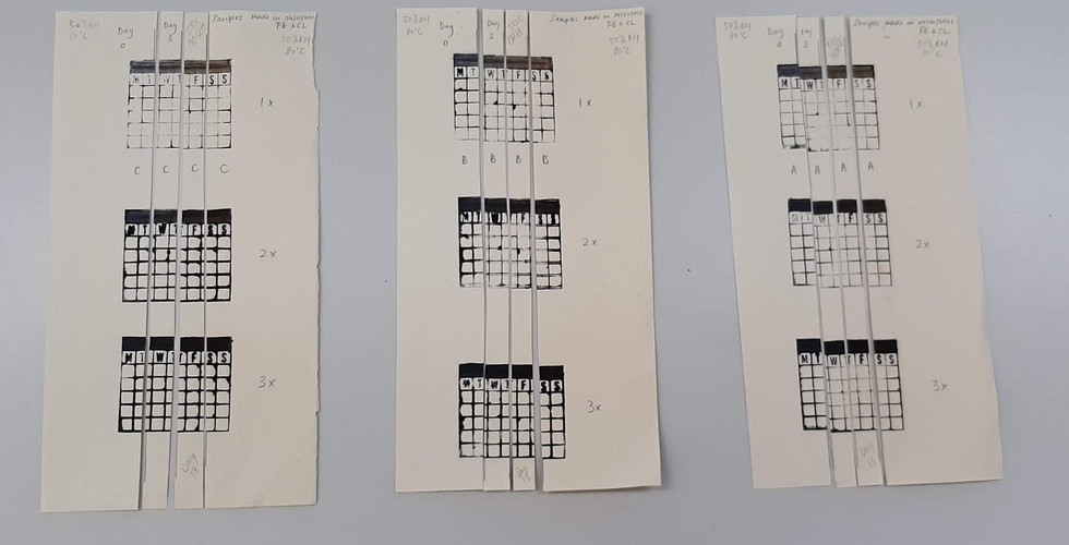
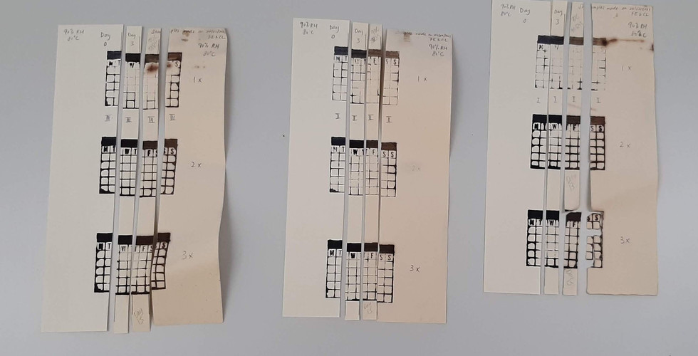
To do this, we decided to try and use stamps to more accurately get a uniform amount of ink in the same area each time. We made 6 samples in total. 3 samples would go into a chamber of 80°C/50%RH, and the other 3 would go into a chamber of 80°C/90%RH.
We cut part of the samples so that of we had a base-line comparison for the progression of degradation. We cut the samples again after 3 days of being in the aging chambers. Already, we saw some degradation. The paper turned slightly yellow and the inks turned slightly brown in all the samples. In the 80°C/90RH samples, we already began to see the iron gall ink becoming more prevalent on the backs of the paper.
Originally, the samples would have been taken out after a total of 10 days. Unfortunately (or fortunately for the experiment's case), the second week of the experimentation was extended due to the class needing to quarantine. For this reason, the sample's progression in the end showed the state of degradation on: Day 0, Day 3, Day 13, Day 17. This led to a large difference between the second and third progression of the sample's degradation but also meant that the final show of degradation is much stronger. While the 80°C/50RH samples do show browning of the paper, some faint bleed-though of the ink, and some blue-brown ink transition, the samples taken from the 80°C/90RH have a significantly higher rate of degradation with sample III showing line breakage and loss of material.


コメント